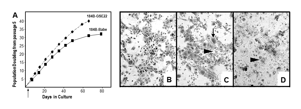
Figure 4. Growth and morphology of post-stasis post-selection 184 with and without functional p53. 184B HMEC were transduced with GSE22-containing or control (Babe) vectors at passage 5. (A) growth curves of 184B-Babe and 184B-GSE22. Note the additional PD in the cultures lacking functional p53. We believe growth rates are similar ± p53, but the absence of p53-mediated growth inhibition allows more cells to continue to proliferate to crisis, leading to apparent faster growth of the population as cells near telomere dysfunction. (B) 184B-Babe at agonescence, 2 months after plating at passage 15, contains mostly large, flat cells with some vacuolization; the cell population can retain this morphology and viability for over a year. (C) 184-GSE22, two weeks after plating at passage 15, shows areas of small proliferating cells and many very large flat cells (arrows). (D) 184B-GSE22, four months after plating at passage 15, shows mostly large multi-nucleated, vacuolated cells and abundant cell debris. All photographs are at the same magnification. (3)